Found in Translation
contributed by Ian Demsky
By its nature, discovery science can be incremental and unsexy — yet it forms the backbone of innovation. Here are just a few of the insights from Rogel Cancer Center labs with the potential to transform clinical care.

A protein involved in cholesterol metabolism plays an important — and previously unknown — role in suppressing the body's natural immune defenders in and around pancreatic tumors, a research team led by the University of Michigan Health Rogel Cancer Center reported in Cancer Research last year.
This discovery of a new player in the pancreatic tumor microenvironment is as typical as it is profound — one of many such nuanced insights into the biology and development of cancer being uncovered across the cancer center’s labs all the time.
Here, the research team found ApoE, an apolipoprotein known to play roles in cardiovascular disease and Alzheimer’s, is elevated in the blood of people with pancreatic adenocarcinoma, with higher levels of ApoE correlating with worse outcomes.
Experiments in mice that lacked the ability to produce ApoE showed reduced tumor growth and more cancer-fighting T cells around the tumors. The researchers also found that ApoE helped drive the production of two immunosuppressive proteins via the low-density lipoprotein, or LDL, receptor, a key player in cholesterol metabolism.
"Not only does our study point to blood levels of ApoE as a possible prognostic biomarker in pancreatic cancer, the findings point toward ApoE as a potential target for improving the effectiveness of chemotherapy and immunotherapy," says study first author Samantha Kemp, Ph.D., who recently graduated from U-M and is now pursuing a postdoctoral fellowship at the University of Pennsylvania. "But there's still a lot we don't know. In pancreatic cancer, it remains to be determined which cell types are targets of ApoE and what functional role it plays in different types of cells."
And thus, insight by insight, our collective understanding of cancer edges forward.
Between the summer of 2020 and the summer of 2021, members of the Rogel Cancer Center contributed to some 250 papers in highly influential journals — sharing new, fundamental discoveries about cancer in places like Nature, Cancer Discovery and Cell.
Years of painstaking laboratory work were added to the world’s pool of knowledge under dry, workaday titles like "Cancer SLC43A2 alters T cell methionine metabolism and histone methylation" and "Loss of optineurin drives cancer immune evasion via palmitoylation-dependent IFNGR1 lysosomal sorting and degradation."
"Biomedical breakthroughs most often rely on a body of discoveries that builds up over time," a recent Nature Cancer editorial noted. "The work that formed the basis of targeted therapies, immunotherapies and the vaccine against human papillomavirus are but a few such examples."
As one of the top institutions in the country for National Institutes of Health funding, U-M is a powerhouse of discovery science research. And the cancer center’s 320-plus members are drawn from nearly 50 departments across campus — ranging from biomedical engineering to surgery, biostatistics to epidemiology, human genetics to medicinal chemistry.
"Basic science research is one of our most important missions — and our members are investigating important areas including the functions of the tumor microenvironment, key signaling changes in cancer cells and tumor stroma, and epigenetic mechanisms of cancer development," says Rogel Cancer Center Director Eric Fearon, M.D., Ph.D., the Emanuel N. Maisel Professor of Oncology. "Yet it's equally important for us to continue to seek opportunities to translate this new knowledge into effective strategies for prevention, earlydiagnosis and treatment."
'A sliver of hope'
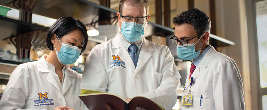
Yoshie Umemura, M.D., and Daniel Wahl, M.D., Ph.D., are a good example of Fearon’s prescription. In 2020, they launched a clinical trial aimed at translating findings from Wahl’s lab that suggested an existing immunosuppression drug may make glioblastomas more vulnerable to radiation therapy.
Despite being the most common type of malignant primary brain tumor, the five-year survival rate for glioblastoma has been stuck below 10%.
"Our main treatments are surgery, chemotherapy and radiation," says Wahl, an assistant professor of radiation oncology. "The standard chemotherapy agent, temozolomide, was approved by the FDA in 2005, when I was just starting medical school — and the needle really hasn’t budged since then."
In most patients, not only do tumors come back after surgery followed by radiation, they grow in the same spot — a hallmark of the disease's inherent resistance, notes Umemura, a clinical assistant professor of neurology.
"That was the genesis of this project," Wahl says. "We wanted to understand what makes these tumors resistant to radiation — and specifically what aspects of the tumors’ metabolism make them resistant."
The research team examined the characteristics of nearly two dozen glioblastoma cell lines, looking at the metabolites they produced and measuring how resistant each was to radiation.
They found that a family of metabolites called purines, which are building blocks of DNA and RNA, correlated with radiation resistance.
"GBM cells that have lots of purines are really resistant to radiation, while GBM cells that have low purines are really sensitive," Wahl says. "And once we had those data we said to ourselves, 'OK, if we intervene and take purines away from the glioblastoma cells, we might be able to make the tumors more sensitive to radiation.'"
Fortuitously, a drug that lowers purine levels had been approved by the Food and Drug Administration two decades earlier. Mycophenolate mofetil, or MMF, had proven safe and effective as an immunosuppressant to prevent organ transplant rejection. It's even available as a generic, lowering the cost of a clinical trial — and potential future treatment costs for patients.
In the lab’s animal model studies, tumor growth was moderately slowed down in mice who received radiation therapy alone or MMF alone, but almost totally halted in the mice who received both. The benefits of MMF were similar whether tumors were grown in the brains of the mice or elsewhere in their bodies, demonstrating the drug’s ability to effectively penetrate the blood-brain barrier — a critical factor for treating patients with brain cancer.
Repurposing a drug that had already been through the rigors of FDA approval smoothed the path between bench and bedside for Umemura and Wahl.
"If we had found a new metabolic enzyme that caused radiation resistance, the reality would be that we’d then have to start doing drug discovery and optimization, maybe spin out a small company, and, in 5-10 years, be ready for a clinical trial — if everything goes well," Wahl notes. "So, yes, one key part was that the drug already exists, and equally important was having a partner like Dr. Umemura who has expertise in developing clinical trials and a university that puts resources behind facilitating this type of translation."
Instead of taking years, the collaboration was able to move forward over a matter of months.
And as the trial proceeded in patients with recurrent glioblastoma, new data from U-M and elsewhere emerged indicating that MMF was indeed safe when given in conjunction with radiation as well as with chemotherapy — and that it made both chemotherapy radiation more effective.
"That gave us the confidence to expand the trial to include patients with newly diagnosed glioblastoma as well," Umemura says. "And last August, they became eligible, too."
She adds, "The bottom line is that we really want better options for our patients. When we’re in an exam room talking to a patient about their prognosis, we want to be able to give them a sliver of hope."
Bedside to bench to bedside
At the Hospital das Clínicas da Faculdade de Medicina at the University of São Paulo, Antonio Lerario, M.D., Ph.D., frequently cared for patients with adrenal cancer. The disease is more common in Brazil than anywhere in the world, especially in children, due to a prevalent TP53 mutation.
Collaborations with the lab of Gary Hammer, M.D., Ph.D., director of the adrenal oncology program at U-M, brought Lerario to Ann Arbor several times as a visiting researcher, and eventually led to him joining Hammer’s lab as a staff scientist.
At U-M, Lerario worked on the effort to map key genomic changes in adrenocortical carcinoma, or ACC, as part of The Cancer Genome Atlas. Michigan was instrumental in having the rare cancer included in the landmark study, and helped assemble the necessary international collaborations to reach critical mass.
"My university, the University of São Paulo, contributed samples to the study and many of them were from patients I used to see in clinic," Lerario says. "So, I knew very intimately the details and history of these patients. That was a motivation for me to come and study these patients' clinical manifestations at a molecular level."
The TCGA project, which was published in 2016, identified three distinct molecular subtypes of adrenal cancer — laying the foundation for the group's continuing efforts to turn those insights into new tests and desperately needed treatments.
"Up to 75% of all patients will eventually develop metastatic disease, and our current medical therapies for ACC provide limited — if any — survival benefit,"
Lerario, Hammer and their co-authors noted in a 2019 commentary.
"While [tumor] grade is certainly effective in pinpointing patients who are less likely to respond to standard of care, such an approach still falls short of rationally directing patients to therapy based on oncogenic pathways driving their specific type of ACC," they wrote.
Building on TCGA insights, Lerario and Dipika Mohan, an M.D./Ph.D. student in Hammer's lab, led the development of an inexpensive, rapid PCR test that boils down a knotty genome-wide signature to a single, yes/no biomarker that can identify whether a patient has the most aggressive of the three flavors of adrenal cancer — with strong potential to guide clinical decision making. (The university has filed for patent protection on the technique.)
More aggressive treatment may be warranted for patients who have the marker because they are unlikely to be helped by mitotane, which is often prescribed after surgery to kill lingering adrenal cancer cells and prevent recurrence, according to retrospective findings the group presented at the 8th International Adrenal Cancer Symposium last fall.
"Of course, guideline-based therapy is best," Mohan says. "But because it’s such a rare cancer, doctors also rely a lot on institutional expertise. These tumors have very high proliferation rates, and we're working with collaborators in Brazil who are using our biomarker assay to try to identify patients who might benefit from earlier cytotoxic chemotherapy — which kills rapidly dividing cells — to shrink the tumors before surgery to help treatment be more effective."
The team is also exploring whether a blood test to capture circulating tumor DNA could be used to classify a patient’s adrenal cancer subtype earlier and non-invasively.
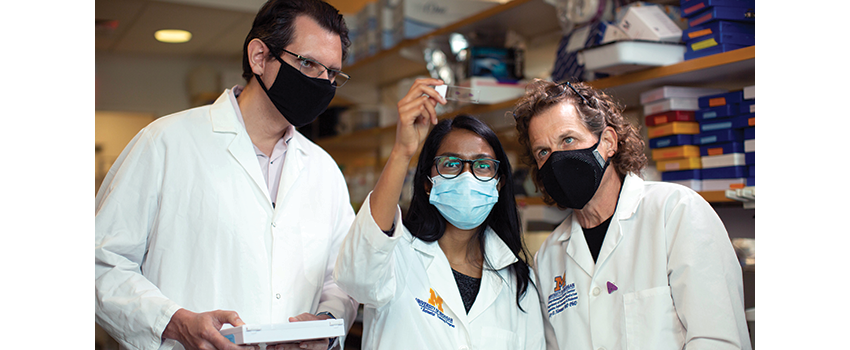
Future tech
Sometimes, innovation means tapping into emerging technologies to improve patient care. Building on their previous research in animal models, Muneesh Tewari, M.D., Ph.D., and Sung Won Choi, M.D., recently teamed up on a small trial that used commercially available, wearable thermometers to detect dangerous complications in hospitalized cancer patients hours earlier than routine monitoring.
The device, which takes readings every two minutes and wirelessly transmits them to the cloud, was able to quickly detect adverse events that affect body temperature, like infection and cytokine release syndrome, in patients who received a hematopoietic stem cell transplant or CAR T-cell therapy — thus allowing for swifter interventions, according to findings their team published in Cancer Cell.
The study also examined more subtle changes in temperature that may appear as deviations from the patient's baseline circadian pattern, signaling a potential problem before a steep temperature rise.
"This additional lead time is clinically significant for patients with cancer, who are commonly immunocompromised and at risk for infection," says Tewari, a professor of internal medicine and of biomedical engineering. "How quickly doctors can administer antibiotics can play an important role in combating potentially fatal infections and sepsis."
The monitoring approach could also facilitate moving some costly inpatient CAR-T care to the outpatient setting by providing additional lead time for the patient to return if a problem was detected, adds Choi, the Edith S. Briskin and Shirley K. Schlafer Foundation Research Professor of Pediatrics. (The study authors and university are pursuing intellectual property protections on the work.)
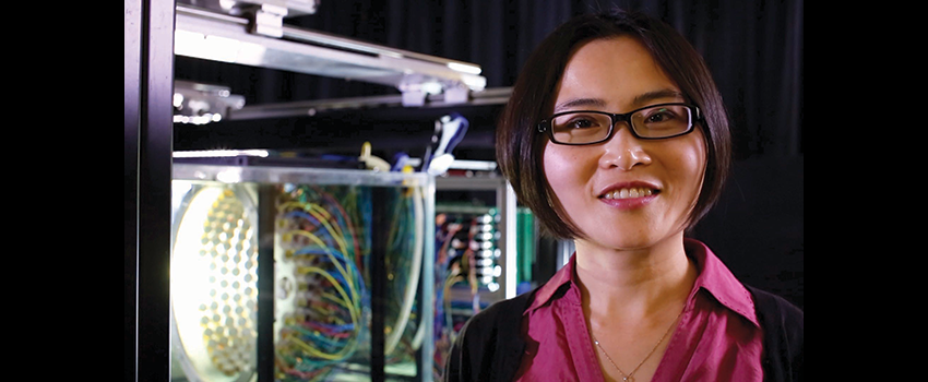
Meanwhile, other cancer center members are pioneering entirely new technologies. Zhen Xu, Ph.D., a professor of biomedical engineering, and a team of U-M of colleagues developed histotripsy, a non-invasive ultrasonic surgical approach.
After nearly 20 years of development, a clinical trial deploying histotripsy against liver cancer recently launched at U-M and seven other sites across the U.S. The trial, which is also being conducted in Europe, is sponsored by HistoSonics, a U-M start-up commercializing the group’s research. (The inventors and university have a financial stake in the company’s success.)
Of course, using ultrasonic pulses is nothing new to medicine, but the process behind histotripsy is. While previous techniques used ultrasound to generate heat energy to ablate tissues, histotripsy harnesses the ultrasound energy to activate thousands of microbubbles at the target — called cavitation — to emulsify tissue. And it can do so with great precision, narrowly targeting the tissues of interest with millimeter accuracy.
"When we started, cavitation was seen as a process to be avoided at all costs," Xu says. "These microbubbles are on the size-order of cells. So, our idea was to use ultrasound to harness and focus this gas we naturally have in our bodies and use these 'micro-scalpels' to target tissue."
The technology is potentially revolutionary on several fronts. It uses an external beam, similar to radiation therapy, which means you don't have to cut into a patient. But more than that, doctors have the advantage of being able to guide the beam using real-time visualization of the target.
"It's non-ionizing, non-toxic and non-thermal," she says. "Huge advantages."
Results from a small, phase 1 trial in liver cancer at three sites in Spain were reported at the February 2020 Society of Interventional Oncology annual meeting. Histotripsy was found to be safe and well-tolerated, and researchers reported the targeted tumors had contracted by 36% after a week, 54% after a month, and 72% after two months.
Additionally, echoing findings from animal studies, in two of the eight patients, tumors other than the ones that were directly targeted also shrank or stabilized following treatment.
This is believed to be a result of a larger immune response triggered by the procedure. Earlier that year, a team led by Xu and co-senior study author Clifford Cho, M.D., a professor of surgery, reported histotripsy ablation stimulated a profound immune response in mouse models of melanoma and liver cancer — and this stimulation of tumor-specific immune response was capable of magnifying the impact of checkpoint inhibition immunotherapy.
"Xu says. "In rodent models, we did several experiments that showed if you treat a portion of the tumor, the remaining tumor can also shrink and disappear, or if you treat one tumor, a distant tumor may also shrink — because the immune system is stimulated to go after more of the cancer."
In June 2021, U-M joined sites around the country in enrolling patients in the #HOPE4LIVER trial, the first large-scale study to evaluate the use of histotripsy against cancer, testing the technology in patients with primary and metastatic liver tumors.
"This research started here, and we are very excited to help move this technology out of the laboratory and into a clinical trial where we can evaluate its safety and efficacy for patients," says interventional radiologist Mishal Mendiratta-Lala, M.D., the principal investigator of the U-M trial site.
Unexpected connections
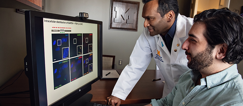
It's often unclear when and how discovery science research might pay off. Serendipity often plays a role both in developing new insights and in applying them outside the lab. Foundational work at U-M uncovered an important gene fusion on-switch for prostate cancer development that has led to the development of a new, sensitive, urine-based prostate cancer test. It also points to new potential prevention and treatment possibilities against COVID-19.
"We weren't exploring this area, but we knew that coronaviruses use two key host proteins to gain entry and replicate within cells," says Arul Chinnaiyan, M.D., Ph.D., director of the Michigan Center for Translational Pathology. "One of those proteins is TMPRSS2. This was appealing because we had discovered TMPRSS2-ETS gene fusions in prostate cancer and knew that TMPRSS2 was regulated by the androgen receptor. It made a lot of sense to explore this in the context of SARS-CoV-2."
Chinnaiyan received a $390,000 grant from the National Cancer Institute to explore the possible role of TMPRSS2 in SARS-CoV-2, and whether treatments commonly used in prostate cancer could potentially be rallied to fight COVID-19.
"The studies we have conducted provide a strong rationale for the use of androgen receptor or BET inhibitors in COVID-19 treatment to decrease TMPRSS2 expression, and that of another key protein, ACE2," he says.
The original TMPRSS2 findings, which were published in Science in 2005, have also led to the development of a urine-based test called MyProstateScore, which is being commercialized by LynxDX, a U-M start-up company. (The inventors and university have a financial stake in the test’s success.)
"The test gives us a non-invasive way to look at a set of exclusive and specific biomarkers to predict whether a patient has an aggressive form of prostate cancer before we move on to an MRI or a biopsy, rather than having everyone with a high prostate-specific antigen level automatically undergo a biopsy," says Chinnaiyan, a co-founder of LynxDx.
A retrospective validation study published in The Journal of Urology found that the MyProstateScore test could have avoided nearly 400 unnecessary biopsies in a cohort of 1,500 patients while failing to flag only 10 aggressive cancers. The test is already available to patients at U-M and the surrounding area.
"There's been a lot of serendipity since the beginning," says Chinnaiyan, the S.P. Hicks Endowed Professor of Pathology and a Howard Hughes Medical Institute investigator.
The initial discovery that the gene fusion underlay 50-60% of all prostate cancers was unexpected, he notes. Gene fusions and translocations were known to be the molecular basis for blood and soft tissue cancers, but virtually unknown in solid tumors at the time.
"It was actually a bit shocking," he notes.
And, of course, back in the early 2000s, there was no way Chinnaiyan and his collaborators could have anticipated that their discovery might have implications in a future pandemic featuring a novel coronavirus that attacks the lungs.
"Still, it’s a homerun hypothesis to pursue," he says. "We have most of the samples we need for this study already banked in our lab. We just never really looked at the lung because there was no reason to at the time. Now we are going back and doing that analysis."
Continue reading the online version of Illuminate Winter 2022 issue:
- The Cancer Microbiome
- Emphasizing Equity
- Perspectives with Max Wicha
- Bringing Harmony to Advanced Breast Cancer Care
- Let's Talk About How We Talk About Race
- Feedback Loops
- Probing a Cancer Paradox
Print/download the Winter, 2022 issue of ILLUMINATE.
Get research news in your inbox!
Our Illuminate e-newsletter showcases the important and unique research underway at the Rogel Cancer Center.
Follow this link and sign-up today!