Researchers are Using Artificial Intelligence to Identify Aggressive Breast Cancer
Media contact: Ian Demsky, 734-764-2220 | Patients may contact Cancer AnswerLine, 800-865-1125
The new approach could help doctors to separate aggressive stage 0 breast cancer from non-aggressive forms, sparing some women unnecessary mastectomies.
About 1 in 5 new breast cancers are caught at their earliest stages, before they’ve spread from milk ducts into the surrounding breast tissue.
But what doctors can’t currently predict with high confidence is which of these cancers — known as ductal carcinoma in situ (DCIS) or stage 0 breast cancer -- are likely to recur and spread after surgery, and which ones surgery is likely to cure.
Researchers at the U-M Rogel Cancer Center have developed a new diagnostic approach using artificial intelligence that aims to do exactly that -- and with greater than 90% accuracy, according to findings published in the American Journal of Physiology-Cell Physiology.
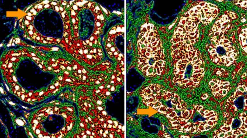
“Scientists don’t really understand what leads to cancer recurrence at the molecular level and that has made it impossible to accurately predict which patients will experience a recurrence and which won’t,” says Howard Petty, Ph.D., a professor of ophthalmology and visual sciences, and of microbiology and immunology at Michigan Medicine, the University of Michigan’s academic medical center. “What we found is that certain key enzymes collect near the cell membrane in these early breast cancers that end up being aggressive, but they don’t in the cancers that are non-aggressive.”
Knowing how aggressive a stage 0 cancer is likely to be could help patients and their doctors decide on the best course of treatment -- which is typically either breast conserving surgery, which consists of removal of the tumor and a small amount of tissue, followed by radiation, or removal of the entire breast.
Importantly, Petty and co-author Alexandra Kraft, now a graduate student in the department of human genetics, found that the abundance of these key proteins didn’t predict a cancer’s aggressiveness, while their location near the cell membrane did.
A critical suggestion that influenced the direction of the research came from an unlikely source -- Petty’s son, a cyber-security expert at a credit monitoring company, who suggested the researchers could use machine learning techniques to teach computers to make increasingly refined connections between subtle features in high-resolution images taken of patient tissue samples and the outcomes those patients experienced.
“The computer is looking for patterns in the images that humans can’t readily discern, from the level of individual pixels up to an entire image of a million pixels,” says Petty, a member of the U-M Rogel Cancer Center.
The researchers started with samples from 70 patients with stage 0 breast cancer who had undergone a mastectomy, and for whom there were at least 10 additional years of medical records available. Twenty of the 70 patients experienced a recurrence of their cancer, while 50 did not.
These tissue samples were stained so that the proteins of interest would fluoresce under the microscope. Then, using a state-of-the-art computer vision application, the scientists created a library of microscope images that were associated either with aggressive or non-aggressive DCIS, based on what had happened to that patient.
Next the researchers showed the program roughly 100 micrographs that it hadn’t seen before -- known as holdout images -- to see how well it could accurately predict whether that patient’s cancer was likely to recur.
With refinements over time, the program is now able to correctly identify aggressive and non-aggressive disease 96% of the time, the team reported.
“That’s pretty impressive when you consider that a human looking at these images would get the answer right about 70% of the time,” Petty says. “And we’ve continued to work on reducing the level of false negatives.”
The program also reported false positives in 4% of cases -- that is, it identified aggressive disease in patients who did not experience recurrence.
“We believe many of these examples speak to the skill of the patient’s surgeon, who effectively cured them of more aggressive disease in the operating room,” Petty says.
As the program continues to be refined using additional samples, Petty is hopeful that with further validation the technology could be approved for clinical use by the Food and Drug Administration within the next few years.
Moreover, he says, the approach could prove effective in predicting the aggressiveness of other similar types of cancer.
“We started with a hypothesis about the biochemical mechanisms that drive cancer recurrence, tested the role of the movement of key proteins to the cell membrane in cancer recurrence and then confirmed our understanding of the underlying biology by assessing how well our explanation predicted what we actually see in patients,” Petty explains. “This improved understanding of the biology of cancer recurrence could also inform the development of new anti-cancer drugs.”
The University of Michigan has filed a patent application on the technology and is assessing options to advance it toward the market.
The research was supported by funding from the Mildred E. Swanson Foundation and the Michigan Economic Development Corporation.
Paper cited: “Spatial locations of certain enzymes and transporters within pre-invasive ductal epithelial cells predict human breast cancer recurrences,” American Journal of Physiology-Cell Physiology. DOI: 10.1152/ajpcell.00280.2020