Circulating biomarkers: Hitting the mark
By Anna Megdell Illustration by Alex Webber
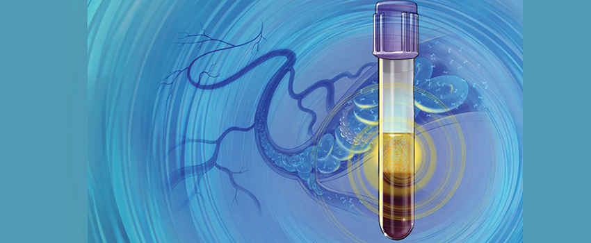
Circulating biomarkers, a new frontier in cancer care, bring both hope and unease to the clinic. Rogel researchers are unraveling their nuances, advancing enabling technologies, advocating for patients and figuring out how to ethically integrate this technology into clinical care.

Imagine a patient comes in for a routine check-up, no symptoms or signs of anything wrong. A blood test reveals markers of early-stage cancer, one that can be cured by surgery.
For a patient receiving chemotherapy, the same type of blood test suggests the cancer is shrinking. In another patient, it points to signs of cancer progressing.
Or, a patient has been told the cancer is removed and they are doing well, but a blood test suggests it’s coming back.
What do the results mean in each situation? How does it influence the next step?
So-called liquid biopsies or circulating biomarkers are a promising beacon for diagnosing cancer or detecting early signs of treatment resistance or metastasis. But researchers’ opinions differ about the effectiveness of the nascent technology—how, and in what settings, can they actually benefit patients?
Circulating tumor biomarkers, found in body fluids such as blood, are proteins, DNA or other substances released by cancer cells, or even cancer cells themselves. These substances or cells come from the cancer tissue and are not seen in normal cells. For solid tumors, the potential of liquid biopsies offers researchers a possible additional tool in their toolbox for measuring treatment response, especially when tumor tissue can’t be easily reached by imaging or standard biopsies.

Traditionally, researchers track tumor shrinkage or growth by measuring the size of the cancer through physical examinations or radiology tests. But what if looking at circulating biomarkers first could tell researchers about the tumor’s responsiveness? If the tumor is growing, researchers might detect more tumor biomarkers in the blood. And if the tumor is shrinking, those same levels may decrease or become undetectable.
Use of circulating tumor markers to track cancer in patients with established metastases, or even in patients who have had cancer in the past and are being monitored, is a well-accepted clinical strategy for many different kinds of malignancies. However, using liquid biopsies to detect a new cancer in a subject without a cancer diagnosis is far from proven. Several recent preliminary studies have suggested that when a blood test demonstrates circulating DNA harboring one or more mutations, it signifies a cancer is lurking in that person.
Since these mutations might come from many different cancers, they have been called multiple cancer early detection, or MCED, tests.
While a lot of hope surrounds this burgeoning methodology of MCED tests, Daniel Hayes, M.D., Stuart B. Padnos Professor of Breast Cancer Research at Rogel, urges colleagues to be honest about the current state of the research.
“Right now, though many researchers and clinical trials are studying it, there aren’t any liquid biopsies proven not just to identify the possibility that the person has cancer, but also to demonstrate that this knowledge helps treat them better,” he says. “We’re just not there yet.”
With this reality, Rogel researchers work to move this promising research forward and clarify this complexity in the clinic.
The Earlier the Better

Courtesy Michigan Medicine
One element of circulating biomarkers that researchers do agree on is in the space of treatment response. Studies indicate that tracking these tumor markers could provide additional data to help determine if therapy is working much sooner than traditional forms of tracking, like CT scans or MRIs.
In a phase 1 clinical trial, Carl Koschmann, M.D., pediatric neuro-oncologist at the University of Michigan Health C.S. Mott Children’s Hospital and clinical research director for the Chad Carr Pediatric Brain Tumor Center, examined cerebrospinal fluid and plasma from patients with diffuse midline gliomas over many months, collecting hundreds of samples through blood draws and lumbar punctures across multiple sites. Koschmann and his team were tracking changes in cell-free tumor DNA (the DNA that circulates in blood and plasma) as glioma patients received treatment concurrent with the clinical trial. Would the cerebrospinal fluid and blood draws hold clues to how the patients were responding to treatment?
The study revealed some promising results. Tumors grew more slowly or took longer to recur in those patients whose allele fraction—the amount of mutant DNA—decreased after receiving the clinical trial drug. This tracked with the researchers’ initial expectations.
But the CSF also revealed something that couldn’t be found in MRI imaging alone: researchers saw a spike in the amount of mutant DNA before the tumor’s growth was captured on the MRI. This was the first study of its kind to collect serial CSF in a clinical trial for gliomas.
“It is very clear that DNA in the CSF can provide a lot of new information about the state of the tumor,” Koschmann says.
Liquid biopsy presents an especially good opportunity for cancers like glioma, where it’s difficult to measure tumor growth and obtaining a traditional tissue biopsy is not feasible.
“If a patient were to come into the clinic right now with a high-grade brain tumor, we’d take an MRI and then make inferences from that imaging about how things are going. But there’s a lot of handwaving about what it means, because it’s the only piece of data we have about how things are going,” Koschmann says. “Patients don’t want to wait until the MRI worsens to change course. Having early information that you might need to adjust treatment is very valuable.”
We Need Both
Sunitha Nagrath, Ph.D., professor of chemical and biomedical engineering, works to develop methods that can isolate, analyze and enable implementation of blood-based biomarkers in the clinic. She says this technology holds tremendous opportunities to monitor patients non-invasively, and to underpin the everchanging molecular phenotypes of a tumor in response to treatment.
“Blood-based biomarkers have changed the landscape of precision medicine. Blood has such a wealth of information, and even with a century of drawing blood routinely for basic patient care in modern medicine, we’ve only scratched the surface. Blood has so much more to offer,” she says.
Muneesh Tewari, M.D., Ph.D., agrees that the research surrounding circulating biomarkers, and the speed with which this area has progressed, holds tremendous hope for the future of clinical cancer care.
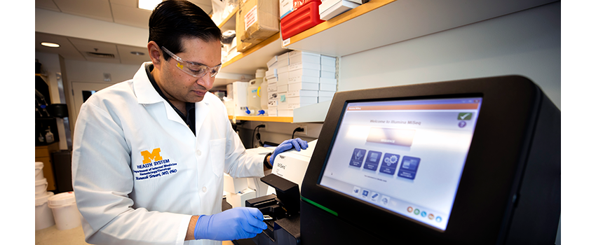
Tewari, Ray and Ruth Anderson-Laurence Sprague Memorial Research Professor, uses next generation sequencing and computational biology techniques to develop biomarker approaches for cancer early detection, disease monitoring and treatment response prediction. A professor of internal medicine and biomedical engineering, Tewari’s lab discovered that in addition to circulating cell-free DNA, RNA could also be found in the bloodstream as a circulating marker, which hadn’t been previously studied extensively.
“Nobody really expected to find them,” Tewari says. “This basic science perspective started my own journey in the field, trying to figure out why circulating RNA biomarkers are there, what cells they are coming from and how they get into the bloodstream.”
When he moved to U-M in 2014, Tewari started studying cell-free tumor DNA in addition to RNA in the blood. He and his team have explored the whole spectrum of circulating biomarkers, from his basic science foundation to translational work and now into improving the technology and application. Broad collaboration is characteristic of this work, given the new and malleable nature of the research.
Tewari works closely with head and neck cancer researchers J. Chad Brenner, Ph.D., and Paul Swiecicki, M.D., to develop better assays in the blood to test biomarkers more effectively. The team has also been developing tools and technology and gathering clinical data to measure cell-free tumor DNA in urine samples as a surrogate for blood samples.
“We’re testing to see whether these blood tests could be converted to urine-based approaches, because urine is completely noninvasive,” Tewari explains.
The idea is that some of the tumor DNA in the blood gets broken down and passes through the kidney’s glomerular barrier. Normally, this barrier prevents large molecules in the blood from getting into the urine, but small fragments of DNA actually do get through.
As someone who has studied circulating biomarkers for over a decade across the spectrum of care, Tewari says that continuing to improve existing tests, developing new tests and investing research into new methodologies is vital because of its profound significance for patients when it comes to early intervention, treatment and recurrence response. Further, the mutations in circulating cell-free DNA may not only reveal that something is going on but also provide insights into specific therapies that might target the mutations found in the DNA.
Fifteen years ago, if somebody with metastatic disease wanted to find out what mutations were present and we didn’t have access to enough primary tissue—something that’s very common in lung cancer, for example—we would have had to do an invasive biopsy,” Tewari says.
Today, he notes, while many patients with metastatic cancer need a biopsy to diagnose the disease, a blood sample can determine which mutations are present and how they’re changing.
“It’s a big deal,” he says. “When we started using the term liquid biopsy in 2006, it was a dream. Now it’s clinical practice from some indications, with many applications on the horizon that are being investigated: early detection of recurrence, monitoring treatment response and ultimately early detection of primary cancer. Each is a huge opportunity for changing patients’ experience and outcomes.”
Despite this hopefulness, Tewari understands the concerns some have around just how new this technology is. He understands it as an inherent part of the process of any kind of medical advancement.
“Of course, there are many issues that need to be ironed out and processes that need to be regulated,” he says. “But there’s always a tension between something new that looks promising and the extent to which you jump forward and start using it, versus validating it and making sure the data is believable. We need both viewpoints. We need to make sure everything is rigorously developed and thoroughly vetted, without which we would have a big mess. At the same time, if we didn’t have some pressure to move things forward and get them into application, it could take decades of just refining and refining, which could be a missed opportunity to benefit patients.”
This is the moment researchers find themselves in: a space where data look promising, and because of commercial pressures, people want to jump ahead. And patients want answers. At the same time, researchers and research-minded physicians understand the necessity to ensure that what is actually being offered to patients is reliable and adds value for patients without causing harm.
“The tension is getting worked through,” Tewari says. “Sometimes it can take years. But eventually, like with any creative process, which research certainly is, that tension will hopefully make the outcome better for patients.”
Part of the Tension
The fulcrum of this issue with the most urgency—in both the potential to improve clinical treatment options and in the wariness of the nascent technology—hinges on its implications for patients and the patient experience.
Some researchers argue that incorporating liquid biopsies into care now is worthwhile given the quicker turnaround of test results for patients. “If someone can know sooner that their tumor has a particular mutation that can be treated with a particular targeted therapy, it can relieve unnecessary stress and anxiety,” Tewari says.
But aside from its ability to track disease progression and recurrence, circulating biomarkers have been touted by companies as a way to detect cancer in otherwise healthy folks, which researchers agree is a breeding ground for unnecessary anxiety for patients.
One of the biggest dangers is false positives, says Elena Stoffel, M.D., M.P.H., clinical associate professor of gastroenterology, who focuses on early detection and cancer prevention.
“All of us have circulating cells that are not normal. Your body’s immune system is in charge of getting rid of cells that are not normal, that are going off program. As we get older, the proportion of cells that are not normal goes up as a normal function of aging. We develop more abnormal cells and many of those abnormal cells may never develop into a cancer, but they may be detectable through some of these biomarker tests,” she says.
The issue lies in the sophistication of liquid biopsy tests used to detect cancer in otherwise healthy patients without symptoms.
“We don’t know what the burden of cells has to be to make one of these liquid biopsy tests positive. We don’t know what the sensitivity or specificity is for these tests. Therefore, it’s possible that entirely by chance, one of the samples might have markers of just a small number of cells that are not normal,” Stoffel says.
The worry is: What do you do when you find that? Where are the cells coming from? What type of cells are they? And if we can tell what type of cells are abnormal, what do we do about that? Do we then go on a diagnostic odyssey involving invasive tests?
“We end up chasing what might be a red herring,” Stoffel says.
Stoffel, who directs Rogel’s Cancer Genetics Clinic, works closely with patients who have greater risk of developing cancer because of genetic mutations that run in their families. For this population, the conversation of circulating biomarkers and liquid biopsies gets even trickier.
“For families at-risk of, say, pancreatic cancer, the recurrent pancreatic cancer screening recommendations are imaging of the pancreas with endoscopic ultrasound or MRI of the pancreas,” she explains. “But there is a biomarker test for pancreatic cancer risk on the market.
It’s not FDA-approved, but it’s on the market. I’ve had several patients ask, ‘Well, should I be doing this in addition to my imaging?’”
Stoffel’s answer? “How is this information going to change anything?”
“I think about a test and the clinical utility of a test by asking, ‘What would I do differently with this information?’” she says. “Because if their imaging is negative and the biomarker test is positive, what are we going to do? Will that information keep the patient up at night? And if the imaging is negative, there isn’t really anything that we can do if the blood test is positive, other than watch and wait—which is what we’re doing anyway with the imaging alone.”
“Another possibility is that the blood test is positive and cancer is found, but maybe that cancer was never going to be clinically evident,” Hayes adds. “In this case, if we do something, is it the right thing for the patient? Given the potential harms of surgery, radiation and systemic therapies for cancers, we need to be sure that a strategy focused on early detection by a liquid biopsy improves a patient’s survival compared to not doing the liquid biopsies.”
When it comes to cancer screening and prevention, Stoffel says it’s important to look to the tests that have proven benefit.
“We know that colon cancer screening and breast cancer screening have proven benefit. But currently, we don’t have any data that shows proven benefit to using a blood-based biomarker test or circulating tumor cells in healthy patients,” she says.
Another concern for Stoffel and Hayes is the risk of false negative tests for those cancers with proven screening methods, like mammography for breast cancer, Pap smears for cervical cancer, colonoscopy for colorectal cancer, high resolution CT scans for lung cancer and more.
“If an MCED test is negative, will patients assume that they don’t need to have these standard screening tests performed?” Hayes says. “We already have evidence that current MCED tests are not always positive when the standard screening tests do show something. It would be terribly unfortunate if the blood MCED tests start to give people a false sense of security. Clearly, nobody likes to have a colonoscopy, but people should not use a negative blood test to forego this lifesaving technology.
“We’re just not there yet.”
In addition to not being FDA-approved, many of the tests on the market that claim to identify disease in otherwise healthy people will require years’ worth of big trials to truly test efficacy, and undertaking those trials, Hayes says, “will require tens, or even hundreds, of thousands of participants and long-term follow-up, which will consume a lot of resources and cost a lot of money.”
For Tewari, optimism about this technology’s ability to detect cancer progression lies in keeping a wide lens.
“Fifteen years ago, any application of these new types of circulating biomarkers was a dream. Today we have tests that are useful at determining what mutations a patient’s cancer has, and ones on the horizon to track recurrence,” he says. “I look at it from that perspective. We just have to manage that tension and do this right—don’t jump forward too quickly but nor should we resist advancement. We need to create and use tests that don’t cause harm and that deliver actual value.
“But in the big picture, I feel like the field is on a good track.”
Continue reading Illuminate, 2023 or download the print version.
Get research news in your inbox!
Our Illuminate e-newsletter showcases the important and unique research underway at the Rogel Cancer Center.
Follow this link and sign-up today!


